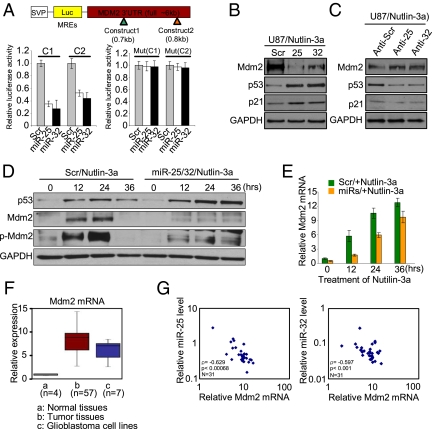
Fig. 4.
Mdm2 is a direct target of miR-25 and -32. (A) Mdm2 3′ UTR contains two predicted miR-25 and -32 binding sites. Reporter constructs, containing a WT (Left) or mutated (Right) Mdm2 3′ UTR, were assayed. (B) Mdm2, p53, and p21 protein levels in U87 at 24 h after transfection with miR-25 and -32 combined with treatment of Nutlin-3a. (C) Mdm2, p53, and p21 protein levels in cells transfected with antisense oligonucleotides against miR-25 and -32 in U87 cells. (D) Mdm2 and p53 expression levels in U87 cells treated with Nutlin-3a after transfected with miR-25 and -32. (E) Mdm2 mRNA expression normalized for GAPDH by qRT-PCR. (F) Mdm2 mRNA relative expression in glioblastoma tissues (n = 57), cell lines (n = 7), and normal brain samples (n = 4) was determined by qRT-PCR assay. The relative expression values were used to design box and whisker plots. (G) Graphic of the negative Spearman correlation coefficient (ρ = −0.629 or −0.597) corresponding to a decreasing monotonic trend between log of Mdm2 mRNA relative expression and log of miR-25 or -32 relative expression (P < 0.00068, n = 31 or P < 0.001, n = 31). (A and E) Data are presented as mean ± SD. We performed three biological experiments in triplicate.

