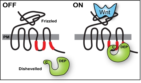
Fig. P1.
Model for Wnt-induced formation of functional Fz–Dvl complexes at the plasma membrane. (Left) Fz receptor in an unbound state. Three Dvl binding motifs in the third loop and cytoplasmic tail of the Fz protein are indicated in red. Dvl resides in the cytoplasm. PM, plasma membrane. (Right) Wnt binding to Fz induces recruitment of Dvl to bind the three-segmented motif in the Fz receptor. Binding involves both the Dvl DEP domain (DEP) and the C terminus (C).

