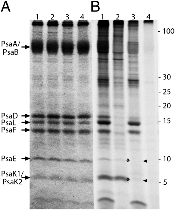
Fig. 3.
Translation of PsaE, PsaK1, and PsaK2 on 80S ribosomes. Equal amounts (45 μg protein per lane) of P. chromatophora PSI labeled in vivo with NaH14CO3 without translation inhibitors (lane 1, A and B), in the presence of chloramphenicol (lane 2, A and B), cycloheximide (lane 3, A and B), or both (lane 4, A and B) were resolved by SDS/PAGE on an 18% polyacrylamide, 7 M urea Schägger gel. Resolved polypeptides were stained with CBB (A) and the 14C signal visualized using a PhosphorImager (B). Asterisks and arrowheads highlight presence or absence (respectively) of radiolabeled PsaE and PsaK1/PsaK2.

