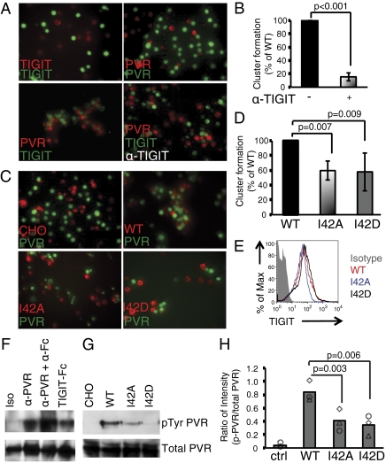
Fig. 4.
Lateral TIGIT/TIGIT homodimerization facilitates PVR signaling. (A and B) Cell-cluster coculture assays performed with stable BJAB cell lines expressing full-length TIGIT or PVR. Cells were labeled with red or green dyes as indicated. (A) Representative images of cell clustering (Upper panels) of cocultured BJAB cells expressing TIGIT or PVR labeled with PKH26 (red) or CFSE (green) and treated with anti-TIGIT antibody as indicated. (B) Quantification of cell-cluster formation for TIGIT-BJAB plus PVR-BJAB cells in the absence or presence of anti-TIGIT antibody by FACS (n = 3). (C and D) Cell-cluster coculture assays performed with stable CHO cell lines expressing full-length TIGIT or TIGIT I42A and TIGIT I42D mutants cocultured with full-length PVR expressed on BJAB cells. (C) Representative images of cell clustering by PVR-BJAB cells with CHO alone, TIGIT-CHO, TIGIT-I42A-CHO, or TIGIT-I42D-CHO. (D) Quantification of CHO-BJAB cell-cluster formation by FACS (n = 3). (E) FACS analysis of cell-surface expression, TIGIT WT (red), TIGIT-I42A (blue), and TIGIT-I42D (black) protein on CHO cells. The gray-shaded histogram represents the isotype control. (F–H) PVR tyrosine phosphorylation assay with iMDDCs. (F) Lysates of iMDDCs—untreated or treated for 5 min at 37 °C with isotype-matched control antibody, anti-PVR, anti-PVR plus anti-IgG, or TIGIT-Fc—were immunoprecipitated with anti-PVR and probed with antibody to phosphorylated tyrosine (α-pTyr; Upper) or α-PVR (Lower). Results are from one of three independent experiments. (G) Human iMDDCs were cultured with CHO, TIGIT-CHO, TIGIT-I42A-CHO, or TIGTI-I42D-CHO cells for 10 min, cell lysates were prepared from isolated iMDDCs, and PVR tyrosine phosphorylation was detected after immunoprecipitation with α-PVR antibody. Representative results from one of three donors are indicated. (H) Quantification of the percentage of tyrosine-phosphorylated PVR of total PVR (with film background subtracted) (n = 3). Each symbol represents iMMDC from one donor.

