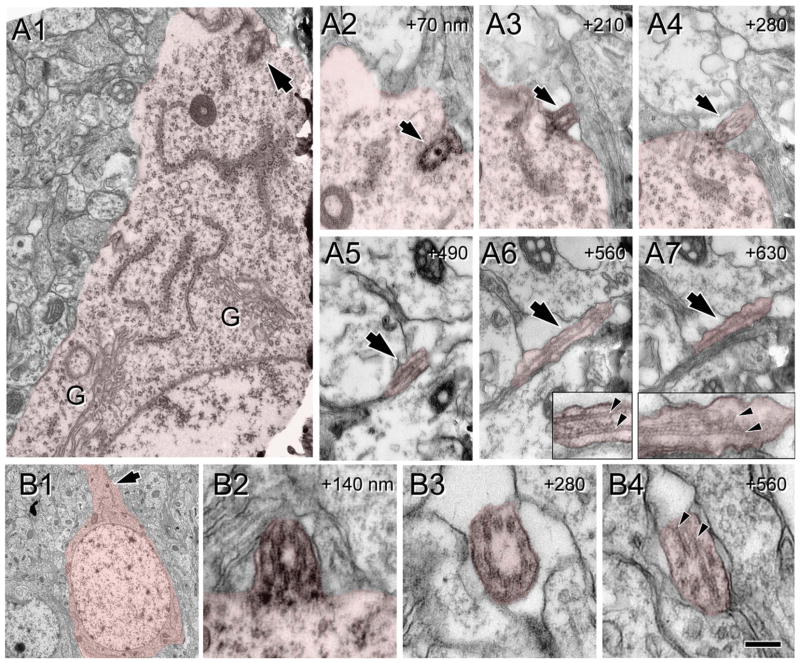
Figure 10.
Ultrastructure of cilia at P8. Cells of interest and procilia are shaded to help with identification. A: Serial micrographs of a cilium at P8. A1: Panoramic view of the cytoplasm and initial apical dendrite of a pyramidal neuron with the basal body attached to the membrane (arrow). G, Golgi apparatus. A2–7: Serial sections of the cilium (arrow) protruding outside of the cell. Well-formed and structured microtubules in the proximal segment (insets in A6,7) appear disorganized distally (inset in A7). Numbers in upper right corner indicate the Z distance between sections in nanometers. B: Serial sections of a cilium from a pyramidal neuron at P8. B1: Panoramic view of the pyramidal cell. Cilium location is indicated (arrow). B2–4: Serial micrographs of the cilia from neuron in B1. B2 illustrates the transition between the basal body and the cilium, and B3,4 are transverse and oblique sections through the cilium showing a straight morphology and a well-developed axoneme along the available length of 0.56 μm. Vesicles were less frequent at this age. Scale bar = 0.12 μm in B4 (applies to B2–4); 0.5 μm for A1; 0.4 μm for A2–7; 5 μm for B1. [Color figure can be viewed in the online issue, which is available at wileyonlinelibrary.com.]

