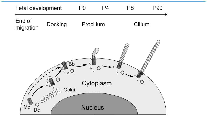
Figure 14.
Model of ciliogenesis stages in mouse neocortical neurons. We propose the following model. Migrating neurons do not bear cilia; rather, their mother centriole (Mc) and daughter centriole (Dc) are free in the cytoplasm. Once cells terminate migration and reach their appropriate lamina, the mother centriole attaches a vesicle, likely from the Golgi apparatus, buds a very short procilium and docks to the plasma membrane. It is also possible that the mother centriole docks directly to the plasma membrane without vesicle attachment as indicated by the dashed arrow. Docking to the membrane involves developing specific structures such as transition filaments, and the mother centriole will become a basal body (Bb), frequently surrounded by vesicles (asterisk). This basal body will grow the procilium, a membranous expansion about 0.5–2 μm in length, whose main feature is the lack of proper axoneme and that typically contains vesicles, short and disorganized tubular structures, and electron-dense diffuse content. This procilium does not display typical axonemal characteristics until ~P8, although axonemal growth seems to start at ~P0 in some subplate cells and could start ~P4 in some populations of neurons that showed early elongation of cilia (e.g., some layer 5 neurons). Overall, cilia will take weeks to elongate fully toward a peak at ~P60–P90, with some differences between layers.

