Figure 1.

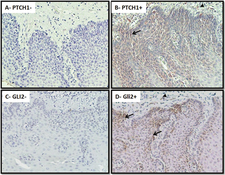
Expression of PTCH1/GLI2 in squamous dysplastic lesions. Expression of PTCH1/GLI2 was examined in 21 cases with squamous dysplastic lesions and 103 cases of ESCCs using standard immunohistochemistry. Summary of our data is shown in Tables S1 and 3. This figure shows representative results. Positives (B for PTCH1 and D for GLI2) and negatives (A for PTCH1 and C for GLI2) staining of severe squamous dysplasia are shown in brown. Please note that expression of PTCH1/GLI2 is mostly in the epithelial lesions (arrows) with some staining in the stroma (arrow heads). Specificities of these antibodies have been shown in our previous publication (see [16] for PTCH1, HHIP; [43] for sFRP1), or by the vendor for GLI2 (Abcam Inc.)
