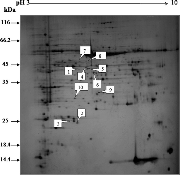
Figure 2.

Representative silver-stained 2D-PAGE gel of separated soybean leaf proteins. Proteins were separated in the first dimension on a nonlinear IPG strip, pH 3.0-10.0, and in the second dimension on a 12% polyacrylamide SDS-gel. Quantitative image analysis revealed a total of 10 spots that changed in abundance.
