
Figure 8.
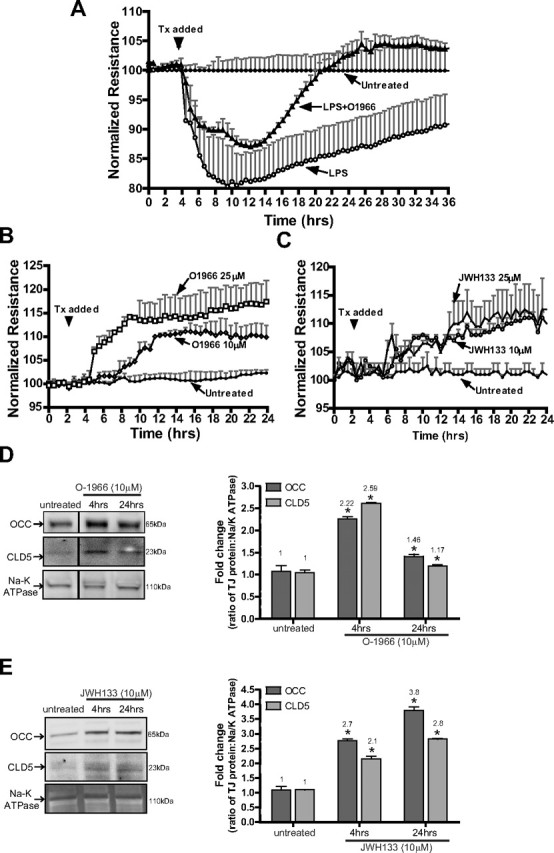
CB2R agonist protects and enhances barrier function in human BMVECs. A, TEER, an indicator of barrier integrity, was measured (by ECIS) in monolayers untreated or treated with either LPS (50 ng/ml) or LPS with O-1966 (10 μm). The resistance was measured at 4 kHz in 30 min intervals for the duration of the time shown. Treatments (Tx) were initiated (arrow) after stable resistance was reached. The data are presented as the percentage change from baseline, which is the resistance measured after treatment divided by the resistance acquired before treatment introduction. B, TEER measurements in BMVECs treated with low (10 μm) and high (25 μm) concentrations of O-1966. C, TEER measurements in BMVECs treated with low (10 μm) and high (25 μm) concentrations of JWH133. CB2R agonists induced a further tightening of the barrier, evident by a rise in the degree of resistance above the basal level (B, C). D, E, Western blots of BMVEC membrane fractions show the tight junction protein levels of occludin (OCC) and claudin-5 (CLD5) in untreated BMVECs and BMVECs treated with O-1966 (D) or JWH133 (E) for 4 h. Densitometry values, indicated to the right, were calculated using the Na/K ATPase loading control, and the data are represented as the fold change from the untreated cells. The asterisk denotes statistical significance (p < 0.05).