
Figure 5. C1 induces the redistribution of Aurora B.
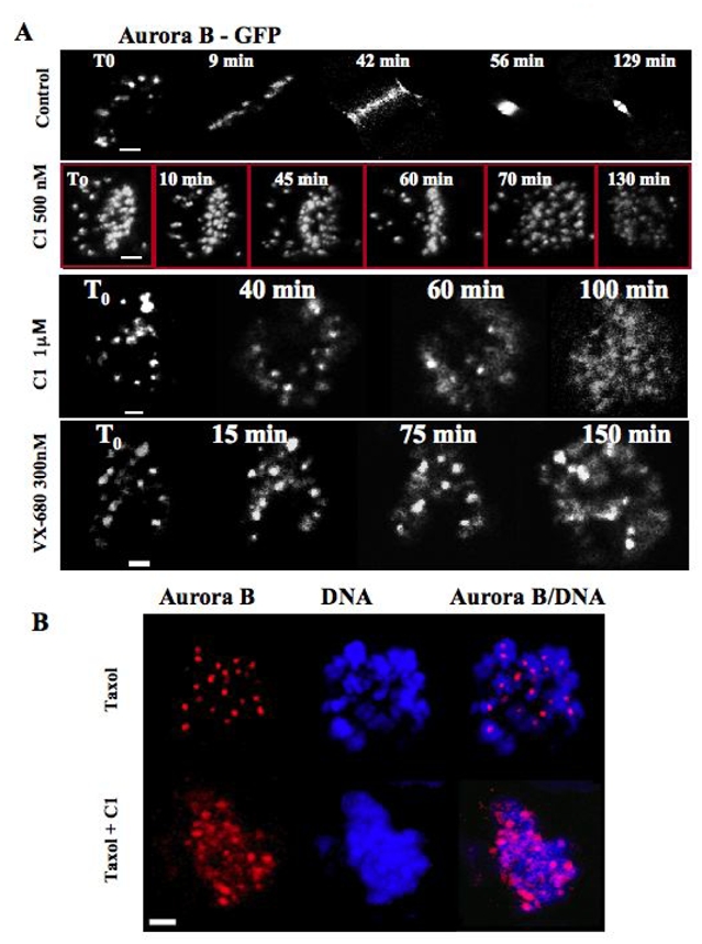
(A) Time lapse microscopy of a mitotic stable HeLa cells expressing Aurora B-GFP fusion in the absence (control) or the presence of either C1 (at 500 nM or 1 μM) or VX-680 (300 nM). The compounds were added to the cell culture and then the behaviour of the cells was continuously imaged. Representative photos, made at the times indicated, are presented. Note that both C1 and VX-680 delayed mitosis onset. In addition, the treatment with 1 μM C1 as well as with VX-680 resulted in a partial delocalization of Aurora B-GFP on the entire chromosomes.
(B) C1 treatment induces also the redistribution of endogenous Aurora B. Overnight paclitaxel (33 nM) treated HeLa cells were incubated for two hours with C1 (1 μM), fixed and analyzed by immunofluorescence microscopy. The localization of Aurora B (red) was detected by anti-Aurora B antibody. DNA (blue) was stained by Hoechst 33342. Note that the strictly punctuated pattern (exclusively centromeric localization) of Aurora B in the control (paxitaxel) cells is no longer observed in the C1 treated cells. Aurora B being partly diffused on chromatin.