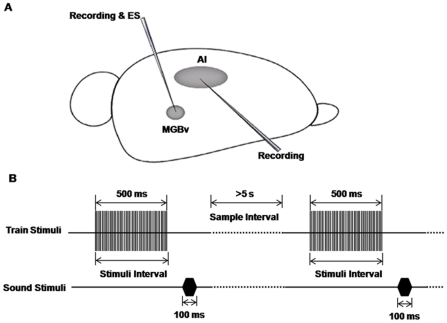
Figure 1. Schematic diagram of experimental configuration and procedure.
A) A tungsten electrode was impelled into the right MGBv to record the electrophysiological characteristics of the neurons in the MGB and verify that the stimulating site was fully within the MGBv. Next, the tungsten electrode was fixed in place to stimulate the MGBv. After that, a sharp glass electrode was impelled to record the intracellular signals of AI neurons. B) Stimulation models. One sample consisted of an electrical stimulus of the MGBv paired with a testing white noise. The intervals between the onsets of the two stimuli were varied from 500 ms to 3000 ms. The intervals between the different samples were longer than 5 s.

