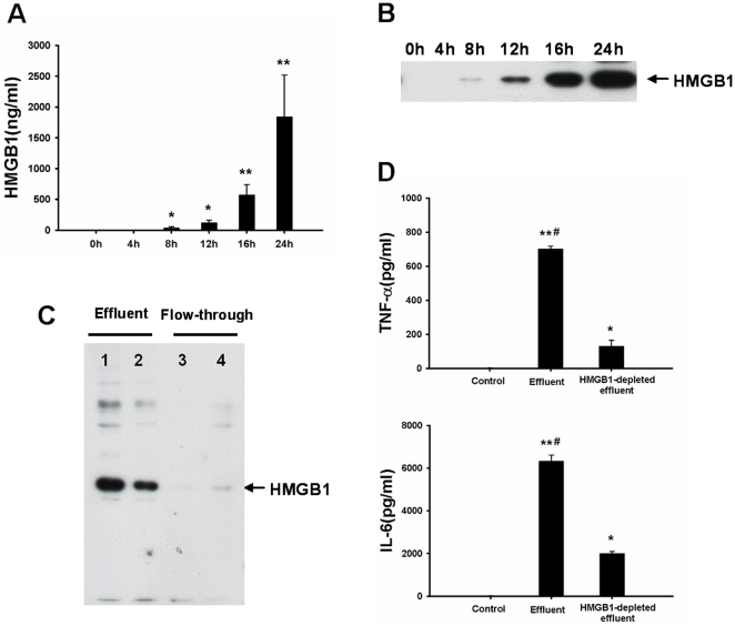
Figure 2. Pro-inflammatory activity of graft effluent is decreased after HMGB1 depletion.
HMGB1 concentration in effluent was detected by ELISA-assay (A) or western blot (B), respectively. Effluent HMGB1 was significantly increased as early as 8 h and then upregulated in a time-dependent manner up to 24 h (*p<0.001, **p<0.0001 vs 0 h). (C) The concentration of HMGB1 in effluent obtained after 24 h of cold storage was drastically reduced after HMGB1 depletion using immunoprecipitation. Lane 1–2 showed the HMGB1 in effluent. Lane 3–4 displayed the HMGB1 in flow-through after HMGB1 depletion. (D) Rat peritoneal macrophages were cultured in the presence of effluent (100 µl), or HMGB1-depleted effluent (flow-through; 100 µl) for 6 h. The inflammatory activity of effluent was markedly decreased after HMGB1 depletion when compared with complete effluent. The experiment was performed in triplicates with similar results. Data are shown as mean ± SD. *p<0.001, **p<0.0001 vs blank control; # p<0.001 vs HMGB1-depleted effluent. HMGB1, high mobility group box 1; TNF-α, tumor necrosis factor-alpha; IL, interleukin.

