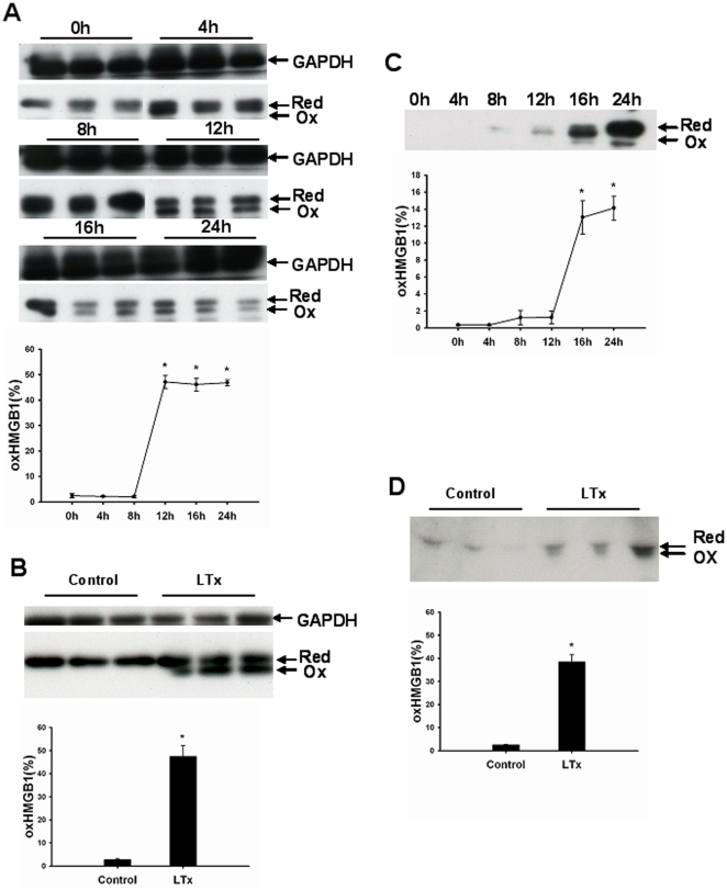
Figure 6. Oxidized HMGB1 is produced after prolonged cold ischemia and reperfusion in liver transplantation.
(A–B) Total hepatic proteins (10 µg) were separated on a non-reducing SDS-PAGE gel and HMGB1 protein was detected by western blot. The gray value of bands was calculated by ImageJ. The relative amount of oxidized HMGB1 was expressed by oxidized HMGB1/total HMGB1. Oxidized HMGB1 was produced in liver graft during cold storage (A) and 24 h after transplantation (B) Each lane represented a separate animal. The results were obtained using liver homogenates from six individual animals per each observation time point. Data are shown as mean ± SD. *p<0.001 vs 0 h. Effluent (C) or serum proteins (D) (10 µl) were separated on a non-reducing SDS-PAGE gel. The gray value of bands was calculated by ImageJ. The relative amount of oxidized HMGB1 was expressed by oxidized HMGB1/total HMGB1. Oxidized HMGB1 was found in effluent after 16 h cold ischemic storage of liver and in serum obtained 24 h after transplantation. The results shown are representative of six animals/group. Data are shown as mean ± SD. *p<0.001 vs normal control rats. HMGB1, high mobility group box 1; GAPDH, glyceraldehyde-3-phosphate dehydrogenase; Ox, oxidized; Red, reduced; Con, normal control; LTx, liver transplantation.

