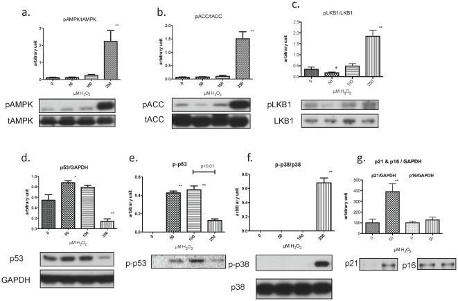
Figure 7. AMPK, LKB1, p53, p21, and p16 protein expression after H2O2 exposure for 1 and 16 hr.
Keratinocytes were treated with the indicated concentration of H2O2 and harvested after 1 hr (a–f) or16 hr (g.) for western blotting (a,b). AMPK activity, assessed by its phosphorylation at Thr172 and that of its substrate ACC Ser79, was increased only at the high (250 µM) concentration of H2O2(c). Phosphorylation of LKB1 (Ser431), an upstream kinase of AMPK, was slightly decreased by 50 µM H2O2 and increased by 250 µM H2O2 (d, e). p53 expression and phosphorylation were increased by 50 and 100 µM H2O2 and were variably affected by 250 µM H2O2 (f). Activation (phosphorylation) of p38-MAPK was only observed with 250 µM H2O2. g. Incubation with H2O2 (50 µM) for 16 h induced the cyclin-dependent kinase inhibitor, p21CIP1 but not p16 INK. (n = 3–6, * p<0.05, ** p<0.01).

