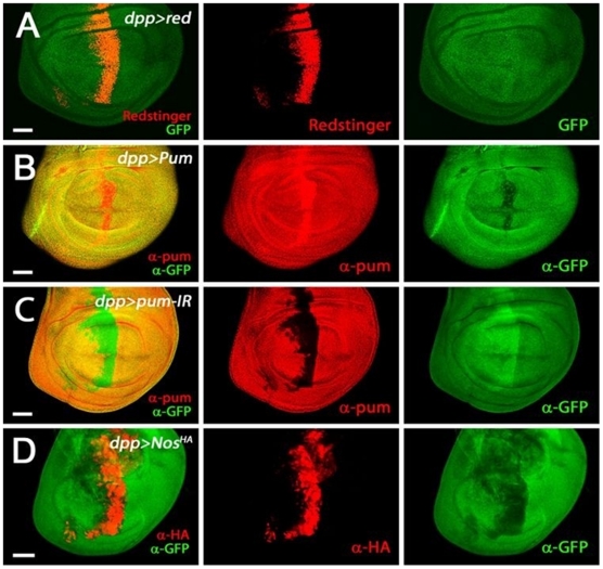
Figure 4. Pum activity in the wing disc.
The 3rd larval wing discs harboring a tub-GFP-NRE construct were visualized by GFP or redstinger fluorescence or immuno-stained by antibodies (α-GFP, α-Pum and α-HA). The dpp-Gal4 driven expression of redstinger (A), Pum (B), Pum-IR (C), and Nos (D) were monitored, as shown in the second column. Pum activity level was monitored by GFP signals, as shown in the third column. The first column is the merged images of the second and third columns. Scale bars indicate 50 µm.

