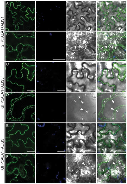
Figure 2. ALA1 localizes to the plasma membrane in the presence of an ALIS protein.
A GFP::ALA1 fusion was transiently expressed in tobacco epidermal cells in the presence of an untagged ALIS. (A,B) ALIS1; (C,D) ALIS3; (E,F) ALIS5. (A,C,E) untreated cells. (B,D,F) cells plasmolyzed in the presence of a concentrated solution of manitol. Arrows in the bright-field and overlay images indicate the relative positions of the plasma membrane and the cell wall in points at which they have detached (opposing arrows) or remain attached (facing arrows) during plasmolysis. The plasma membrane is indicated with a closed arrow and the cell wall with open arrows. Green: GFP fluorescence; blue: autofluorescence; gray: bright field image. Panels on the right show an overlay image. Scale bar: 25 µm.

