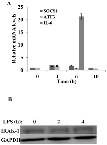
Figure 5. Socs1 and Atf3, negative regulators of TLR4 signaling, are not induced and IRAK-1 remains intact upon LPS stimulation in kidney fibroblasts.
(A) The expressions of Socs1 and Atf3 were not induced after stimulation with LPS. Wild-type kidney fibroblasts were either untreated or treated with 100 ng/mL LPS for 4, 6, or 10 hours. Socs1, Atf3, and Il-6 transcripts were measured by real time RT-PCR assays and standardized against Gapdh levels. Each experiment was performed in triplicate. Data is depicted as means +/− standard deviation. (B) LPS does not cause the degradation of IRAK-1. Wild-type kidney fibroblasts were either untreated or treated with 100 ng/mL LPS for either 2 or 4 hours. Whole cell lysates were harvested and analyzed by Western blot with IRAK-1 specific antibodies.

