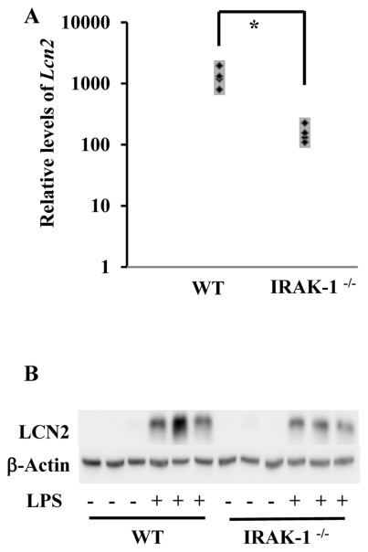
Figure 6. Loss of IRAK-1 causes decreased expression of Lcn2 mRNA and protein in vivo.
WT and IRAK-1−/− C57/BL/6 female mice of 12 weeks old (6 each) were intraperitoneally injected with either 30 mg/kg of LPS or PBS for 6 hours. (A) Kidney tissues were extracted and subject to RNA extraction. Lcn2 transcripts were measured by qRT-PCR assays and standardized against their respective controls (mice receiving PBS injections). Data is depicted as three separate mice (both WT and IRAK-1−/−). *P<0.05 (B) Protein lysates were extracted from the kidney tissues and subjected to Western blot. Blots were analyzed using LCN2 specific antibodies. The same blot was probed with β-actin as a loading control.

