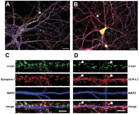
Figure 1. Subcellular localization of a-syn and UCH-L1 in mature cultured hippocampal neurons.
Representative images of hippocampal neurons immunolabeled with anti-a-syn, anta-synapsin I and anti-MAP2 antibodies (A, C) or anti-a-syn, anti-UCH-L1 and anti-MAP2 antibodies are shown (B, D). The straightened dendrites in (C) and (D) correspond to the regions indicated by arrows in the whole cell images (A, B). Arrowheads in (D) indicate selected regions where UCH-L1 colocalizes with a-syn. Representative max z-projected confocal images (cell and straightened dendrites) are depicted. Whole cell (A, B) scale bar = 20 µm; dendrite (C, D) scale bar = 5 µm.

