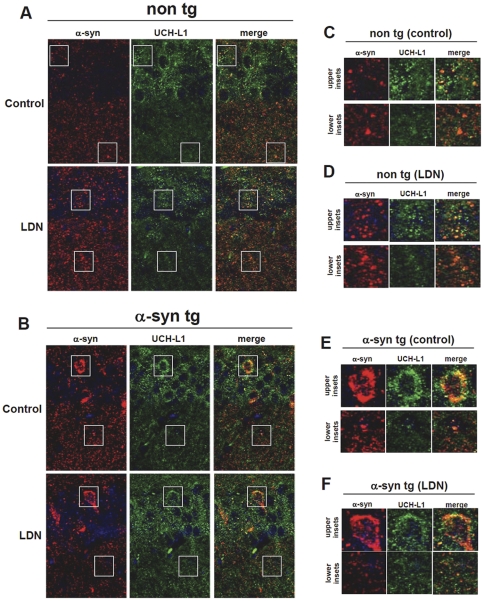
Figure 5. Subcellular localization of a-syn and UCH-L1 in non tg and a-syn tg hippocampal sections.
Representative hippocampal vibratome sections from control and LDN-treated non tg (A) and a-syn tg (B) mice that were double-immunolabeled with antibodies against UCH-L1 and a-syn. The antibody against a-synuclein recognizes both human and mouse forms of this protein. Samples were further stained with DAPI to visualize nuclei (blue). Magnified inset correspond to boxed areas from non tg control (C), non tg LDN-treated (D), a-syn tg control (E), and a-syn tg LDN-treated (F) are shown. The upper insets correspond to the upper boxed regions and the lower insets correspond to the lower boxed regions in each representative image.

