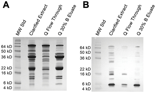
Figure 2. SDS-PAGE and immunoblot analysis of anion-exchanged purified lunasin.
Aliquots of samples corresponding to the bench-scale anion-exchange chromatography method where lunasin was eluted using a step gradient (Figure 1C) were subjected to SDS-PAGE and immunoblot analysis. (A) SDS-PAGE of the clarified extract, column flow through (Q flow through), and the 30% Buffer B elution (Q 30% B Fraction). Clarified extract, Q flow through, and Q-30%B fraction were prepared at dilutions of 1∶8, 1∶8, and 1∶10, respectively, and electrophoresed using 15% Tris-glycine gels. Molecular weight standards (MW Std) are shown in the first lane. (B) Immunoblot analysis of the clarified extract, Q flow through, and the Q 30% B Fraction. Proteins were separated by SDS-PAGE as described in (A), transferred to a PVDF membrane, and probed with a lunasin-specific mouse monoclonal antibody. For SDS-PAGE, clarified extract, Q flow through, and Q-30% B fraction were prepared at dilutions of 1∶20, 1∶20, and 1∶40, respectively. Molecular weight standards (MW Std) are shown in the first lane.

