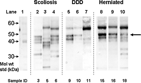
Fig. 1.
Identification of biglycan core protein in IVD via Western blotting. The intact core protein can be seen for most samples at 45 kDa (large arrow). Bands of lower molecular weight represent fragments of biglycan core protein down to 25 kDa in size, sometimes with multiple fragments. In some samples an intense band was observed at 55 kDa, which may be non-specific or cross-reactive with PRELP (see text). Details of individual samples can be seen in Table 1. DDD degenerative disc disease

