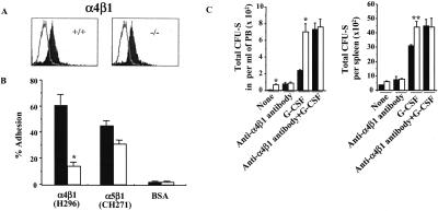
Figure 2.
Effect of Rac2 deficiency on integrin-mediated cell adhesion. (A) Expression of integrin α4β1 (VLA-4) analyzed by flow, one representative experiment of three with identical results, with biotin-conjugated anti-α4 mAb. The solid curve represents expression of α4β1 as detected by mAb; open tracing represents an isotype control. (B) Adhesion of HSC/P cells to FN H296, containing the heparin binding site and the CS-1 binding site for α4β1, or to FN CH271 containing the heparin binding site and the RGDS binding site for α5β1. Closed bars, WT mice; open bars, Rac2−/− mice. Mean ± SD, *, P < 0.01. (C) Enumeration of CFU-S12 in peripheral blood after treatment with G-CSF alone or with both G-CSF and anti-α4β1 antibody. Closed bars, WT mice; open bars, Rac2−/− mice. Mean ± SD; *, P < 0.01.

