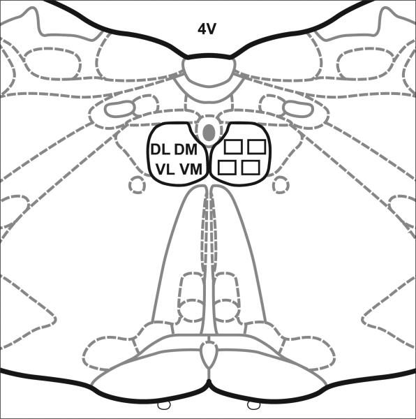
Figure 1.
The location of sample areas in the hypoglossal nucleus. The paired hypoglossal nuclei were divided into quadrants. One image was taken from each quadrant: dorsolateral (DL), dorsomedial (DM), ventrolateral (VL) and ventromedial (VM) on each side. 4V, fourth ventricle. Diagram modified from (Paxinos & Watson, 1998).

