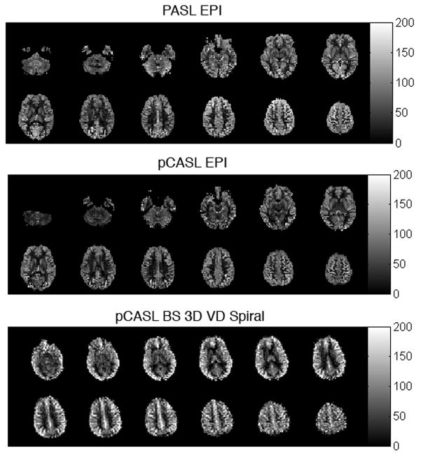
Figure 1.
Typical whole-brain ASL MRI quantitative CBF data obtained in 6 minutes at 3 Tesla using PASL and pCASL with echoplanar imaging (TOP and MIDDLE), adapted from (109) with permission from the publisher. BELOW: pCASL with background-suppressed 3-dimensional variable density spiral acquisition acquired in 2 minutes at 3 Tesla.

