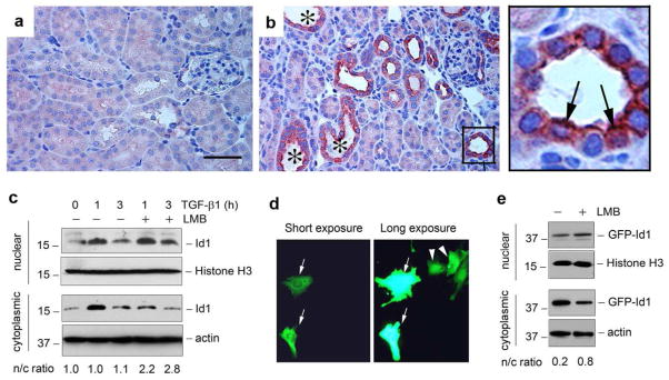
Figure 2. Id1 is induced in both cytoplasm and nuclei of renal tubular epithelial cells in the injured kidneys.
(a, b) Immunohistochemical staining shows Id1 induction in the degenerated tubules after obstructive injury. (a), sham. (b), UUO for 7 days. Id1 induction was observed in the degenerated tubules with dilated lumen (*). Boxed area in Panel (b) was enlarged. Arrows indicate the tubular epithelial cells with both cytoplasmic and nuclear staining for Id1. Scale bar, 50 μm. (c) Western blot analyses demonstrate the presence of both cytoplasmic and nuclear Id1 protein. Human kidney tubular epithelial cells (HKC-8) were treated with or without TGF-β1 (2 ng/ml) in the absence or presence of leptomycin B (LMB) as indicated. Cytoplasmic and nuclear protein were prepared and immunoblotted with antibodies against Id1, histone H3 or actin, respectively. The ratio of nuclear/cytoplasmic Id1 (n/c ratio) was calculated and presented in the bottom of Panel (c). (d) Subcellular localization of the GFP-Id1 fusion protein. HKC-8 cells were transfected with GFP-Id1 expression vector. Two images in the same area with different exposure times are shown. Arrows show the cytoplasmic localization of GFP-Id1, while arrowheads denote a predominant nuclear location. (e) Nucleocytoplasmic shuttling of GFP-Id1 fusion protein. HKC-8 cells were transfected with GFP-Id1 expression vector, followed by incubation with or without LMB as indicated. Cytoplasmic and nuclear proteins were immunoblotted with different antibodies as indicated. The ratio of nuclear/cytoplasmic Id1 (n/c ratio) was given in the bottom of Panel (e).

