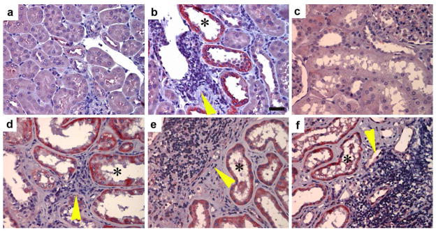
Figure 3. Id1 induction is closely associated with peri-tubular inflammation in chronic kidney diseases.
(a, b) Immunohistochemical staining shows a selective upregulation of Id1 in the degenerated, dilated tubules in mouse model of diabetic nephropathy. (a), control kidney; (b) diabetic kidney. Id1 was selectively induced in the dilated tubules (*) in the proximity of a cluster of inflammatory cells (yellow arrowhead). Scale bar, 25 μm. (c–f) Id1 is induced in renal tubules of kidney biopsies from patients with different nephropathies. (c), control kidney; (d) focal and segmental glomerulosclerosis (FSGS); (e, f) diabetic nephropathy. Asterisks (*) denote the Id1-positive renal tubules. Yellow arrowheads show the infiltration of inflammatory cells.

