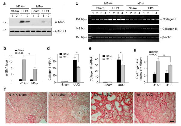
Figure 9. Id1 deficiency attenuates myofibroblast activation, inhibits matrix gene expression and ameliorates fibrotic lesions in obstructive nephropathy.
(a, b) Western blot demonstrates that deficiency of Id1 inhibited α-SMA expression in the obstructed kidneys after UUO. Kidney lysates were immunoblotted with specific antibodies against α-SMA and GAPDH, respectively. Representative Western blot (a) and quantitative data of α-SMA expression in various groups (b) are presented. *P < 0.05 (n = 6). (c–e) Id1 deficiency inhibits matrix gene expression n obstructive nephropathy. (c) representative RT-PCR analysis of renal mRNA levels of type I and type III collagen in different groups as indicated. Numbers denote each individual mouse in a given group; (d, e) quantitative determination of renal type I (d) and type III collagen (e) mRNA levels by real-time PCR in various groups as indicated. *P < 0.05 (n = 6). (f) Representative micrographs show the deposition of collagen as detected by Picrosirius Red staining. Bar = 25 μm. (g) Renal total collagen content was estimated by measuring hydroxyproline levels in obstructed kidneys at days after UUO. Hydroxyproline levels were expressed as μg/mg dry kidney weight. *P < 0.05 (n = 6).

