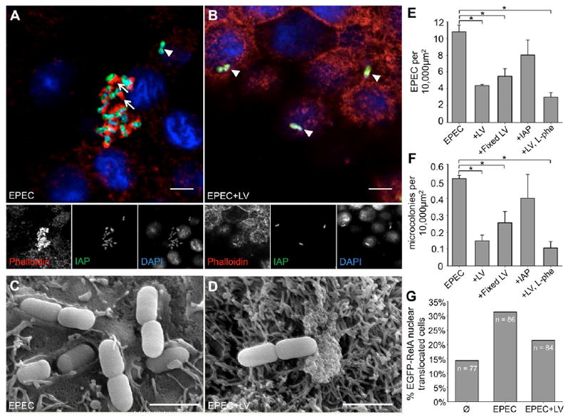
Figure 2. LVs prevent EPEC attachment to IECs.

(A, B) CM images of HT-29 monolayers treated with EPEC ± LVs stained for IAP (green), F-actin (phalloidin, red) and bacteria (DAPI, blue), show striking enrichment of IAP surrounding attached bacteria. Most EPEC in non-LV treated samples (A) form micro-colonies and are associated with actin pedestals (arrows), indicating intimate attachment, whereas this rarely occurs in the presence of LVs (B). Arrowheads in A, B denote examples of EPEC superficially attached to the monolayer, indicated by the absence of pedestal formation. Bars, 5 μm. (C, D) Representative SEM images of CACO-2BBE cells demonstrate EPEC intimately associated with the cell surface in the absence (C), but not the presence of LVs (D). Bars, 1.67 μm. (E) The number of EPEC attached to HT-29 cells is significantly reduced in the presence of LVs, LVs fixed in paraformaldehyde, and LVs with IAP activity inhibited by L-Phe, but not in the presence of purified IAP (*p < 0.05). (F) LVs also reduce the number of micro-colonies formed. (G) Nuclear translocation of EGFP-RelA is increased in HT-29 cells incubated with EPEC, an effect which is partially ameliorated by treatment with LVs.
