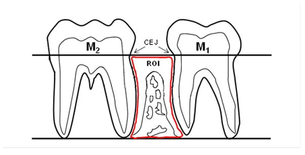
Figure 1.

Schematic view of the region of interest (ROI) for the quantitative histomorphometric analysis. Histomorphometric analyses were performed within an area located at the interproximal alveolar bone between the first mandibular molar (M1) and the second mandibular molar (M2). The ROI boundaries are: coronally - a longitudinal line projected through the cementoenamel junction (CEJ) of M1 and M2; apically - a longitudinal line at the level of the apices of the roots of these molars; and mesially and distally - the distal root surface of M1 and the mesial root surface of M2, respectively.
