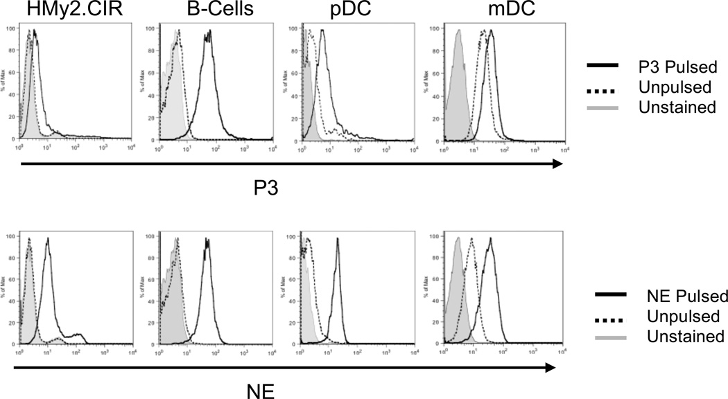
Figure 2. NE and P3 are taken up by APCs.
HMy2.CIR cells and healthy donor APCs, including B-cells, pDCs and mDCs, were pulsed with P3 (5 µg/mL) (top panel) or NE (5 µg/mL) (lower panel) for 3 hours, permeabilized and stained with FITC-conjugated antibodies to NE and P3, and then analyzed using flow cytometry. Unpulsed mDCs demonstrate baseline staining for NE and P3 as they endogenously express both proteins.

