Figure 1.
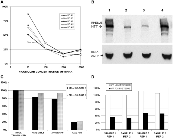
(A) Level of rhesus huntingtin messenger RNA expression (normalized to GAPDH) in LLC-MK2 cells transfected with various concentrations of candidate anti-huntingtin small interfering RNA (siRNA), relative to levels in mock transfected cells. (B) Western blot of rhesus Huntingtin (HTT) and beta-actin proteins in lysates from LLC-MK2 cells transfected with anti-huntingtin small interfering RNA candidate HD1 or HD5. Lanes: (1) untreated cells, (2) cells transfected with HD1, (3) cells transfected with HD5, (4) cells transfected with a scrambled control small interfering RNA. (C) Level of huntingtin messenger RNA expression in HEK293T cells transduced with AAV2-CTRL5, AAV2-GFP or AAV2-HD5, relative to levels in mock transduced cells. Data from two separate cell experiments are shown. (D) Level of rhesus huntingtin messenger RNA expression in striatal brain tissue from two samples of GFP-positive versus two samples of GFP-negative cells obtained by laser capture microdissection from the pilot monkey co-infused with AAV2-HD5 and AAV2-GFP. Data from two replicate assays are shown.

