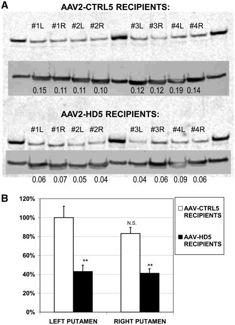
Figure 6.
(A) Western blot of Huntingtin protein in tissue punches from left (L) and right (R) putamen of the four monkeys receiving AAV-CTRL5 (top) or the four receiving AAV-HD5 (bottom) Lanes 2–5 and 7–10 (Lanes 1, 6 and 11 are protein standards). Insets show same blot re-probed and imaged (at a shorter exposure time) for tubulin protein. Numbers in each lane provide the ratio of Huntingtin to tubulin densitometry values. (B) Amount of Huntingtin protein (normalized to tubulin) in left or right putamen of each group of monkeys, relative to average amount in left putamen of monkeys receiving AAV-CTRL5, **P < 0.01 AAV-HD5 versus AAV-CTRL5 by hemisphere. NS = left versus right putamen difference in AAV-CTRL5 recipients is not statistically significant.

