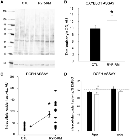
Figure 3.
Oxidative stress and increased oxidative markers in RYR1-related myopathy (RYR1-RM) myotubes. (A, B) Increased oxidative stress markers as measured using oxyblot assay. (A) Representative oxyblot from control (CTL) and RYR1-related myopathy myotubes. (B) Quantitation of mean total carbonyls showing a significant increase in RYR1-related myopathy myotubes as compared to control (*P < 0.001). (C) Basal oxidant activity expressed in arbitrary units (AU) was measured by use of the DCFH assay in myotubes from RYR1-related myopathy (n = 8) and control subjects (n = 4). A significant increase of 69 ± 20% was observed in patient myotubes (asterisks). Each dot represents the average value of the fluorescence intensity of 10 myotubes. (D) Quantitation of oxidant activity using DCFH assay from control (black) and RYR-RM (white) myotubes after incubation with apocynin (Apo, n = 19) or indomethacin (indo, n = 19). Results are reported as per cent of oxidant activity compared with DMSO treated myotubes. A small but significant decrease was observed in RYR1-related myopathy myotubes after apocynin treatment (#P < 0.001).

