Figure 1.
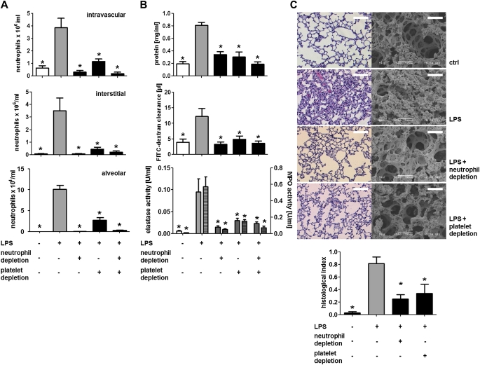
Neutrophils and platelets cooperate in the onset of LPS-induced acute lung injury. Mice were challenged with LPS by inhalation and killed 4 hours later. Neutrophils were depleted by injection of an anti-Ly6G antibody (clone 1A8, 50 ng, intraperitoneally), whereas platelets were depleted by application of antiplatelet serum (50 μl, intraperitoneally). (A) Quantification of intravascular (top), interstitial (middle), and alveolar neutrophils (bottom). (B) Protein concentration (top), fluorescein isothiocyanate (FITC)–dextran clearance (middle), and elastase (bottom, uniform bars) and myeloperoxidase (MPO) activity (bottom, hatched bars) in bronchoalveolar lavage fluids. (C) Representative histologic (left) and scanning electron microscopic (right) images of lungs from mice treated as indicated. Scale bar indicates 50 μm for scanning electron microscopy and 250 μm for histology. Quantification of histologic lung sections is shown at bottom. n = 8–10 for each bar. Statistical significance was tested using one-way analysis of variance with Dunnett post hoc test. *Indicates significant difference compared with LPS-treated animals.

