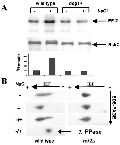
Figure 4.
EF-2 phosphorylation in vitro and in vivo. (A) In vitro kinase assay with Rck2 and EF-2 as substrate. C-terminally HA epitope-tagged Rck2 from wild type and a hog1 deletion strain was immunoprecipitated before (−) and after (+) induction by osmotic shock as described in Materials and Methods. The amounts of precipitated Rck2 kinase were tested by Western blotting, and incorporation of 32P was quantified with a PhosphorImager (Molecular Dynamics). (B) Two-dimension gel analysis of EF-2 modification in vivo. Protein extracts were prepared before (−) and 15 min after osmotic shock (+) and separated on two-dimensional gels as described in Materials and Methods; (−/+) mixture of both extracts; + λ PPase, mixture treated with λ phosphatase. EF-2 was detected with polyclonal antibodies.

