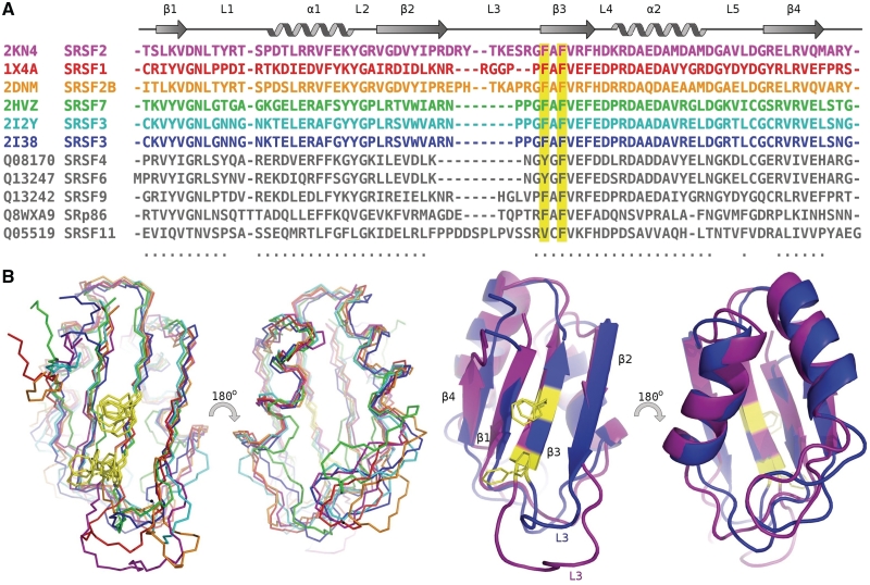
Figure 2.
Alignment of the SR protein RRM domains. (A) PDB deposited structures colored according to overlay and identified by PDB number and name, all other RRMs colored grey and identified by uniprot number and name. Conserved residues for RNA binding are highlighted in yellow (B) Left: Overlay of backbone atoms of the molecular structures of all known SR RRM domains, backbone alignment using 57 residues (indicated in by dots beneath the sequence) with an RMSD of 1.18 Å. Structures shown of human SR family RRM domains; SRSF1 (1X4A–RSGI), SRSF2B (2DNM–RSGI), SRSF7 [2HVZ (16)] and SRSF3 [2I2Y, 2I38 (16)]. Right: Cartoon representation of structures; for clarity only two SR–RRM domains are shown; SRSF2 and SRSF3, the SRSF3 structure used for this alignment is from PDB ID 2I2Y, the only SR–RRM structure determined in the presence of RNA.

