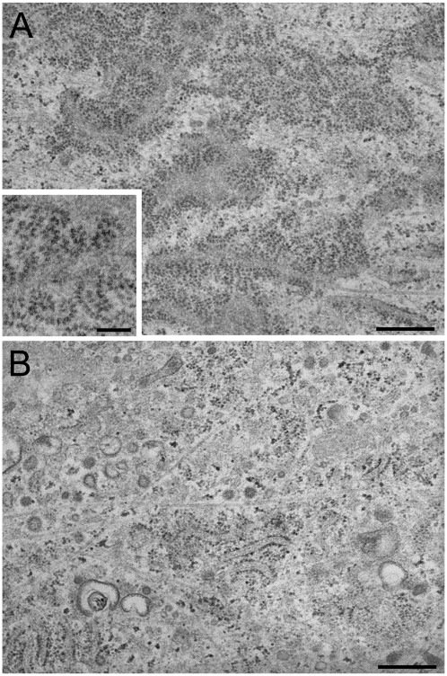
Figure 1.
Transmission EM images of the rough ER surface in collagen-secreting HEL fibroblasts. Control (A) and p180 siRNA-transfected (B) HEL cells were grown in F12/DMEM containing 2% FBS in the presence of ascorbate. A large number of highly developed polysomes are clearly seen on the membrane surface in ascorbate-stimulated HEL fibroblasts, while p180 depletion results in dramatically reduced numbers of polysomes on the ER. Polysomes are frequently seen in the membrane surface view when the sections were cut parallel to the substrate. A higher magnification image of control cells (inset in A) shows spiral arrays of polysomes consisting of about 25–30 ribosomes. Bars: A and B, 500 nm: inset in A, 200 nm.

