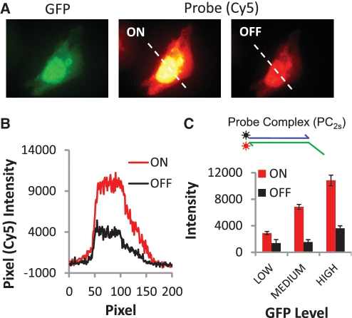
Figure 1.
Labeling and removal of Cy5 fluorophores from protein markers via strand displacement reactions of a two-strand probe complex (PC2s). (A) Selective labeling of expressed GFP proteins in CHO cells. The images display a strong correspondence between the GFP and two-strand probe (Cy5) signals; pixel intensities of the GFP and Cy5 signals are linearly correlated (r2 = 0.94). However, OFF signal intensities indicate ∼20% of active Cy5 dye remains on the cells after the erasing reaction. (B) Pixel intensities for cross section indicated in the probe images in A for both the ON and OFF states of the cells. (C) Histogram of the average Cy5 signal intensities for the ON and OFF states of 20 cells. Cells are grouped based on their GFP whole-cell fluorescence intensities (low, medium and high levels correspond to 2000–5500, 5500–10 000 and 10 000–15 800 intensity units, respectively).

