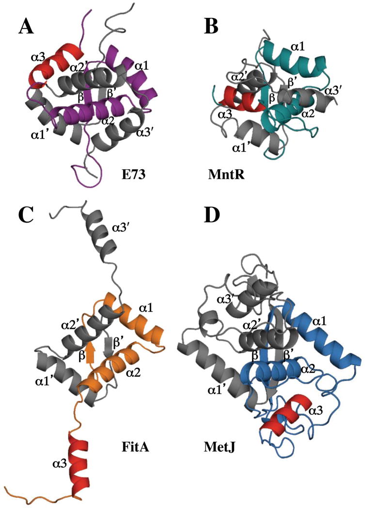
Figure 6.
Comparison of the structure of E73 (A) with that of other RHH proteins containing a 3rd helix including (B) MntR (PDB ID:1MNT); (C) FitA (PDB ID: 2BSQ); and (D) MetJ (PDB ID: 1MJM). All structures are shown as ribbon representations with one protomer colored in grey and the other colored in purple (E73), green (MntR), orange (FitA), or blue (MetJ). The 3rd helix of the colored protomers is shown in red for all 4 proteins to highlight that the orientation of the 3rd helix in E73 relative to its RHH motif is unique to E73 and that all three homologs have very distinct 3rd helix topology.

