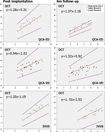
Fig. 7.
Passing-Bablok non-parametric orthogonal regression of minimal lumen area (MLA) measured by the different imaging modalities (x axis) and that measured by optical coherence tomography (y axis). Data immediately post-implantation and at 6 months follow-up. ED edge detection, IVUS intravascular ultrasound, MLA minimal lumen area, OCT optical coherence tomography, QCA quantitative coronary angiography, VD videodensitometry

