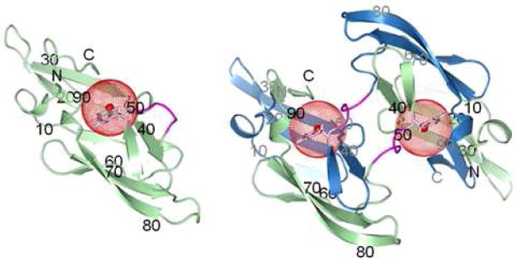
Figure 1.

Structures of wt CV-N monomer (left, PDB ID: 2EZM)27 and domain-swapped dimer (right, PDB ID: 3EZM).29 Ribbon diagrams are shown with chains A and B colored in green and blue, respectively, and the hinge-loop in magenta. The side chain of W49 is shown in stick representation (pink) with a red sphere of radius 5 Å drawn around the fluorine atom at position 5 of the tryptophan ring. Amino acid sequence positions are labeled for every 10th residue, in black for chain A and in gray for chain B.
