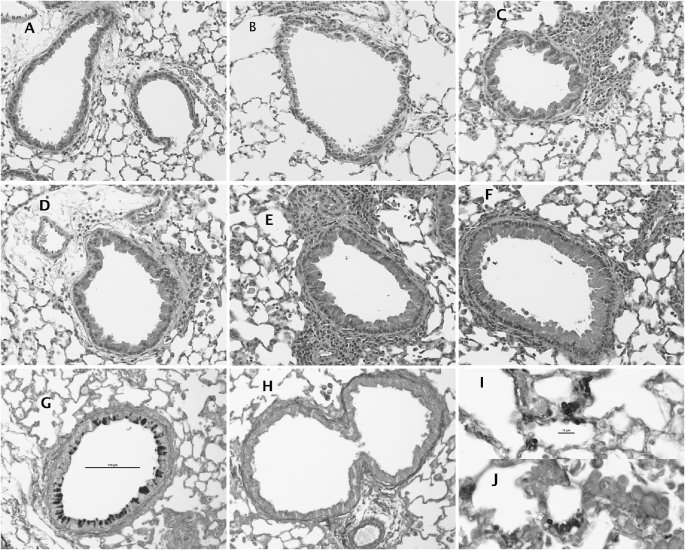
Figure 5.
Histologic appearance of pulmonary tissue (A and B) before, and (C, D, G, and H) 4 and (E, F, I, and J) 7 days after Pneumocystis inoculation in (A, C, E, G, and I) BALB/c mice and (B, D, F, H, and J) C57BL/6 mice. Stain is standard hematoxylin and eosin in A–F, Gomori silver stain in G–J. Total magnification: ×200 (except for I and J: ×600).

