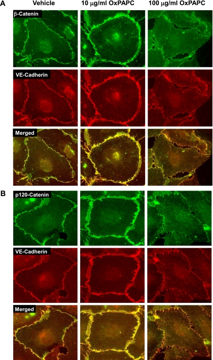
Figure 3.
Immunolocalization of p120-catenin and β-catenin in OxPAPC-stimulated ECs. ECs grown on glass coverslips were stimulated with OxPAPC (10 μg/ml or 100 μg/ml, for 30 minutes), followed by double immunofluorescence staining for (A) β-catenin (green) and VE-cadherin (red), and (B) p120-catenin (green) and VE-cadherin (red). Merged images depict areas of protein colocalization, which appear in yellow. The results shown are representative of three independent experiments.

