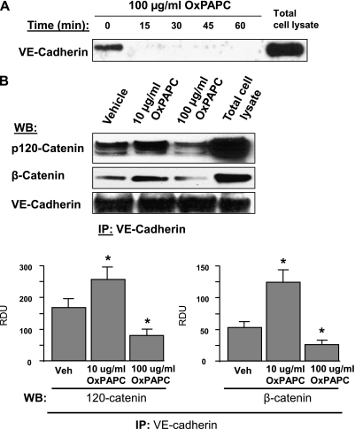
Figure 5.
Effects of OxPAPC on VE-cadherin internalization and interaction with β-catenin and p120-catenin. (A) Cells were stimulated with OxPAPC for indicated periods of time and washed, and cell-surface proteins were labeled with Sulfo-NHS-SS-Biotin, as described in Materials and Methods. Cells were lysed, and biotinylated proteins were precipitated with streptavidin–agarose. The presence of biotinylated VE-cadherin was evaluated by Western blot analysis. (B) VE-cadherin was immunoprecipitated from cells stimulated with OxPAPC (10 μg/ml or 100 μg/ml, 30 minutes). Amounts of coimmunoprecipitated p120-catenin and β-catenin were evaluated by Western blot analysis. The bar graph depicts results of quantitative analyses of VE-cadherin–p120-catenin and VE-cadherin–β-catenin associations. All experiments were repeated three times. Values are mean ± SD. *P < 0.05, versus control samples. IP, immunoprecipitation; Veh, vehicle; WB, western blot.

