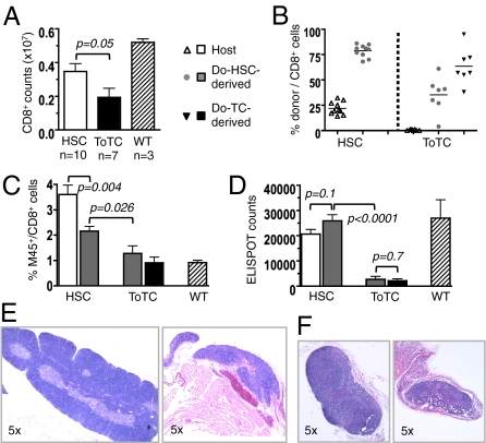
Fig. 2.
Late CD8+ cell recovery and response to MCMV. BALB.B recipients of B6 HSCs or B6 HSC+ToTCs were infected 8 wk after HCT with a sublethal dose (1 × 10⁵ pfu) of MCMV RM427+. WT controls were similarly infected. Tissues were harvested 2 wk postinfection. (A) Higher absolute CD8+ T-cell numbers per spleen in HSC vs. HSC+ToTC recipients. (B) FACS chimerism analysis of spleens revealed mixed donor/host CD8+-chimerism in HSC recipients; the CD8+ pool in HSC+ToTC recipients comprised primarily cotransferred donor cells, few HSC-derived donor cells, and no host cells. (C) In HSC recipients, the percentage of M45-tetramer+ cells was higher among residual host cells compared with donor HSCs. In recipients of HSC+ToTCs, the cotransferred CD8+ T-cell pool had the lowest percentage of M45-tetramer+ cells, and their HSC-derived donor CD8+ cells had lower percentages of M45 reactivity vs. HSC-derived CD8+ cells in HSC recipients. (D) ELISPOT analysis of FACS-sorted donor and host populations. In HSC recipients, ELISPOT activity of donor and host-derived CD8+ cells was equivalent to that in WT controls. HSC+ToTC recipients had significantly lower IFN-γ responses. (E) H&E staining (magnification of 5×) of thymuses of representative HSC (Left) and HSC+ToTC (Right) recipients 8 wk post-HCT showed normal morphology for HSC recipients, but atrophy, hypocellularity, and loss of follicular structure for HSC+ToTC recipients. (F) H&E staining (magnification of 5×) of peripheral lymph nodes showed normal morphology for HSC recipients (Left), but decreased size, atrophy, hypocellularity, and disrupted morphology in HSC+ToTC recipients (Right) at 8 wk post-HCT.

