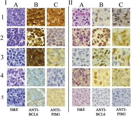
Fig. 4.
Concurrent expression of PIM1 and BCL6 in lymphomas, mouse and human, B- and T-cell, by immunohistochemistry. I, Representative murine lymphomas. In each case, histologic analysis shows malignant tumor cells (H&E stain in column A). 1–3, BCL6-positive lymphomas (brown nuclei, B) from BCL6 transgenic mice containing viral insertions within or near the PIM1 gene (first two rows are T-cell tumors as demonstrated by flow cytometry; Fig. 1B), and row 3 is a B-cell tumor (Fig. 1A). PIM1 levels are positive in all these lymphomas (brown nuclei, C), and the level of staining correlates roughly with the intensity of BCL6 expression. Rows 4 and 5 are lymphomas from control mice (row 4 is T-cell, row 5 is B-cell); BCL6 (B) and PIM1 staining (C) are negative in both controls. II, Representative randomly selected human lymphomas. Histology analysis indicates malignant neoplasm in each case (A, H&E stain). 1–3, Randomly chosen BCL6-positive B-cell tumors previously studied for diagnostic purposes. 4, Randomly selected BCL6-positive lymphoma previously classified diagnostically as T-cell. BCL6 and PIM1 staining are positive (brown nuclei) in each case. 5, Previously studied T-cell lymphoma that does not stain for BCL6 or PIM1.

