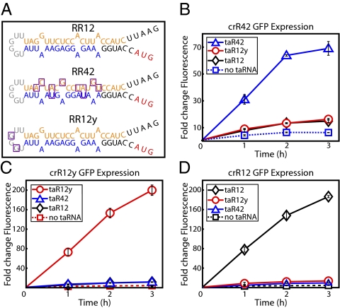
Fig. 2.
Characterization of riboregulators RR42 and RR12y. (A) Mfold-predicted secondary structures of crR12, crR42, and crR12y. Mutations in variants crR42 and crR12y, relative to the parent crR12 variant, are outlined in purple. Important features are color-coded: cis-repressive sequence (orange), RBS (blue), target gene start codon (red), and taRNA recognition bases (gray). (B–D) Fold changes in GFP expression from fully induced crRNA cotranscribed with cognate and noncognate taRNA. (B) crR42. (C) crR12y. (D) crR12. All values were normalized by OFF state (no crRNA or taRNA induction). Graphs depict the triplicate mean ± SEM.

