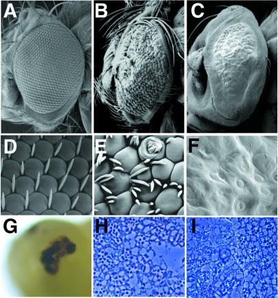
Figure 1.
Phenotypes of neur mutant eyes. (A–F) Scanning electron micrographs (SEM) of adult eyes; (A–C) ≈×150; (D–F) ≈×5,000. (A, D) Wild-type eyes. (B, E) neurA101 mutant eyes generated with ey-FLP. Note tufting of interommatidial bristles, irregular sizes of ommatidia, pitting, and scarring of ommatidia. A strong degree of head macrochaetae tufting around the perimeter of the eye is also apparent. (C, F) neurIF65 mutant eyes generated with ey-FLP. Eyes are bald and smooth; ommatidia lack definition and lenses. Note that head macrochaetae are also absent. (G) Pupal head from an animal aged 45 h after puparium formation containing neurIF65 mutant eyes; a large necrotic patch at the position of the developing eye is present. (H, I) Tangential plastic sections through a neurA101 mutant eye generated by using ey-FLP (H) and an eye containing a neurIF65 mutant clone generated by using hs-FLP (I, mutant clone is left of the dotted line). neurA101 ommatidia frequently have too many rhadomeres, whereas neurIF65 clones have extremely disrupted rhabdomere morphology and numbers; compare with the normal stereotyped arrangement of rhabdomeres in wild-type ommatidia (I, right of the dotted line).

