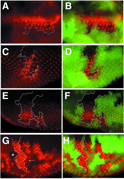
Figure 2.
neur is required for lateral inhibition of photoreceptors. Clones of neurIF65 (A–F) and neurA101 (G, H) were generated with hs-FLP and are marked by the absence of nuclearly localized GFP detected in green; selected clone boundaries are outlined in white. The expression of different antigens detected in red (A, C, E, G) are shown merged with the GFP clonal marker (B, D, F, H). (A, B) Ato expression normally resolves to single presumptive R8 cells but remains expressed in large clusters within the clone and persists longer than in neighboring wild-type cells. (C, D) Boss is normally expressed in single differentiated R8 cells, but large clusters of Boss-positive cells are found within the clone. Different focal planes are shown in C and D because Boss is apically localized and the clone marker is nuclearly localized; this leads to a slight displacement in the positions of the mutant clone and the phenotypically mutant cells. (E, F) Elav is present in all photoreceptors. A large excess in Elav-positive cells is present within the clone. (G, H) Expression of β-galactosidase in neurA101 homozygous clones. A cell-autonomous increase in the number of β-galactosidase-positive cells is observed within mutant clones; compare with neighboring heterozygous tissue (G, arrow). Note that mutant cells are also homozygous for the enhancer trap and thus produce more β-galactosidase per cell than heterozygous tissue or twin-spot clones, which produce none (G, star).

